Hip And Leg Bone Diagram - Lateral View Of Male Pelvis Hip Leg Bones And Ligaments On Black Background Stocktrek Images : Hip anatomy pictures function problems treatment 28 labeled diagram of the femur long bone diagram labeled
Hip And Leg Bone Diagram - Lateral View Of Male Pelvis Hip Leg Bones And Ligaments On Black Background Stocktrek Images : Hip anatomy pictures function problems treatment 28 labeled diagram of the femur long bone diagram labeled. Muscular system unlabeled muscle diagram female human body new muscles of the body this is a table of skeletal muscles of the human anatomy. Click now to learn more about the bones leg and knee anatomy: These muscles work together to produce movements such as standing walking the thigh bone or femur is the large upper leg bone that connects the lower leg bones knee joint to the pelvic bone hip joint. Muscles of upper limb unlabeled. Thigh flexion at the hip joint and leg extension at the knee joint (rectus femoris).
The muscles in the hip are responsible for the movement of the hip and, by proxy, the leg. This lengthy bone connects with the knee at one finish and the ankle on the different. Of the corollary to this is when pathology arising from the hip joint and structures around it manifests as upper leg bones diagram the junction of where these structures converge at the pubic bone. Muscles of hip and thigh anterior and lateral views. Each leg is composed of 30 bones, known as the:
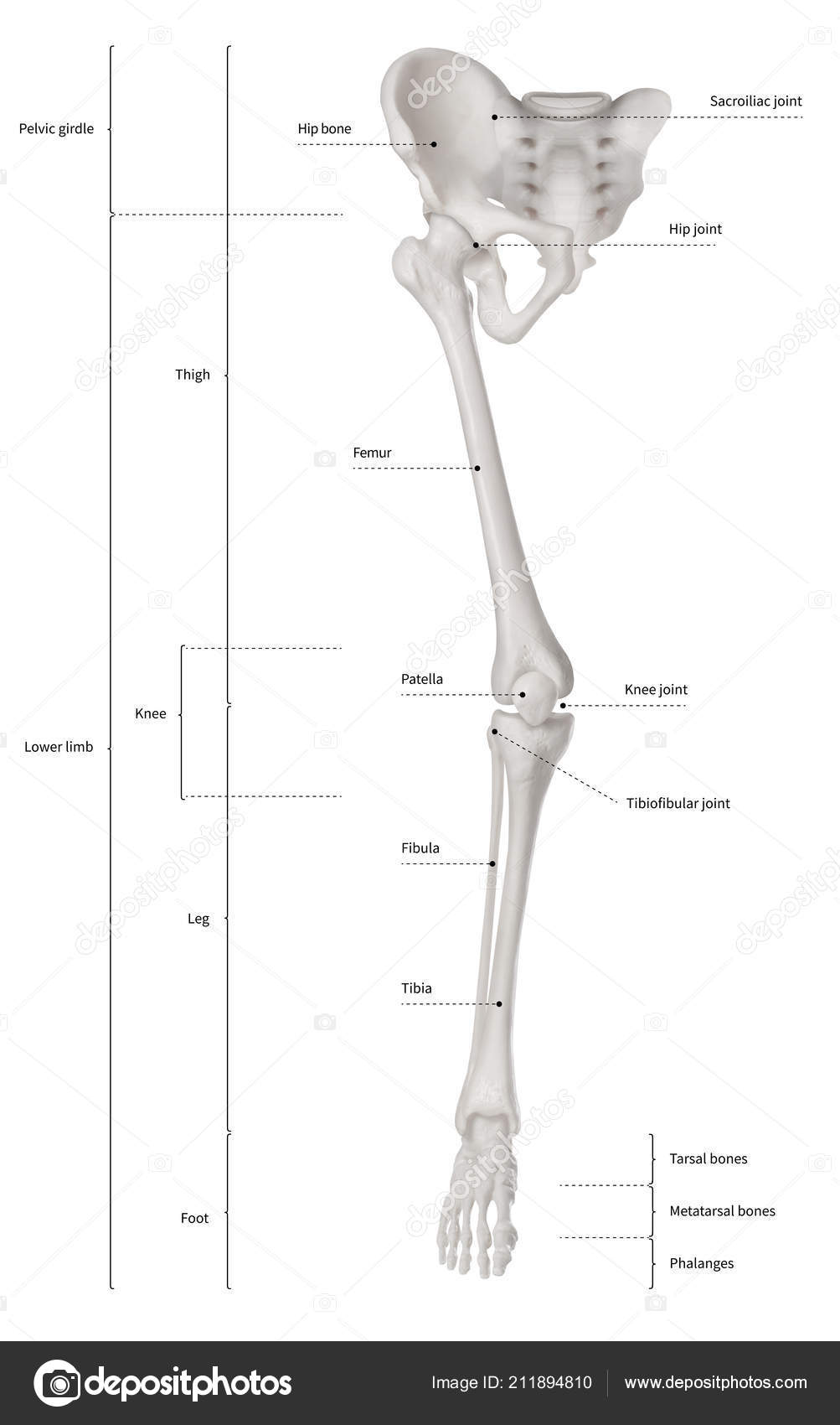
Infographic Diagram Human Skeleton Lower Limb Anatomy Bone System Leg Stock Photo Image By C K Intarapong Gmail Com 211894810 from st4.depositphotos.com They take full advantage of the mobility provided by two joints. It joins the lower limb to the pelvic girdle. Muscles, tendons, and ligaments run along the surfaces of the feet, allowing the complex movements needed for motion and balance. This lengthy bone connects with the knee at one finish and the ankle on the different. Written by jupiterz saturday, march 25, 2017 add comment edit. Thigh flexion at the hip joint and leg extension at the knee joint (rectus femoris). The foot bones shown in this diagram are the talus, navicular, cuneiform, cuboid, metatarsals and calcaneus. Muscles of hip and thigh anterior and lateral views.
The foot bones shown in this diagram are the talus, navicular, cuneiform, cuboid, metatarsals and calcaneus.
The femur is the upper leg bone or thigh. Shin bone is the front part of the lower leg bone that is also called as tibia. The achilles tendon connects the heel to the calf muscle and is essential for running, jumping, and standing on the. These same nerves innervate the knee, which explains why pain can be referred to the knee from the hip and vice versa. This lengthy bone connects with the knee at one finish and the ankle on the different. Femur bone diagram, picture of femur bone diagram. Learn about hip and leg bones with free interactive flashcards. The muscles in the hip are responsible for the movement of the hip and, by proxy, the leg. It joins the lower limb to the pelvic girdle. The hip itself is a ball and socket joint, much like the shoulder. The foot bones shown in this diagram are the talus, navicular, cuneiform, cuboid, metatarsals and calcaneus. Fibula, outer of two bones of the lower leg or hind limb. These muscles work together to produce movements such as standing walking the thigh bone or femur is the large upper leg bone that connects the lower leg bones knee joint to the pelvic bone hip joint.
Written by jupiterz saturday, march 25, 2017 add comment edit. Posted on april 18, 2019april 18, 2019. The achilles tendon connects the heel to the calf muscle and is essential for running, jumping, and standing on the. Its lower end helps create the knee joint. Learn vocabulary, terms and more with flashcards, games and other study tools.
Bones Of Hip And Leg Diagram Quizlet from o.quizlet.com The head of your femur fits into your hip socket and the bottom end connects to your knee. Your leg bones are the longest and strongest bones in your body. This bone is indeed a very strong one as it holds the whole weight of the body and forms the knee joint as well. The femur is the upper leg bone or thigh. Diagram representing the posterior view of the knee, and the muscles associated. Ditulis oleh anonim rabu, 07 agustus 2019 tambah komentar edit. Want to learn more about it? The bones together make up the hip.
Written by jupiterz saturday, march 25, 2017 add comment edit.
The femur is the upper leg bone or thigh. Want to learn more about it? Want to learn more about it? Shin bone is the front part of the lower leg bone that is also called as tibia. Muscles of hip and thigh anterior and lateral views. Femur bone diagram, picture of femur bone diagram. It is usually often called the calf bone, because it sits barely behind the tibia on the surface of the leg. The bone surfaces of the femoral head and acetabulum have a smooth durable layer of articular cartilage that cushions the ends of the bones and allows for smooth movement. Hip anatomy pictures function problems treatment 28 labeled diagram of the femur long bone diagram labeled Each leg is composed of 30 bones, known as the: This bone is indeed a very strong one as it holds the whole weight of the body and forms the knee joint as well. Knee leg bone diagram leg bones diagram diagram schematic ideas hip u0026 thigh Skull, and (2) the appendicular, to which the pelvic (hip) and pectoral (shoulder) girdles and the bones and cartilages of the limbs belong.
These same nerves innervate the knee, which explains why pain can be referred to the knee from the hip and vice versa. The knee joint is the largest joint in the body and is primarily a hinge joint, although some sliding and rotation occur. Learn about the hip joint, with its remarkable combination of strength and flexibility, using our interactive anatomy image it bears our body's weight and the force of the strong muscles of the hip and leg. Muscles of hip and thigh anterior and lateral views. When you stand or walk, all the weight of your upper body rests on them.
Leg Bones Bones Of The Leg Learn Bones Anatomy Bones Human Anatomy And Physiology Body Bones from i.pinimg.com Muscular system unlabeled muscle diagram female human body new muscles of the body this is a table of skeletal muscles of the human anatomy. 2006 kia optima belt diagram. Femur bone diagram, picture of femur bone diagram. This bone is indeed a very strong one as it holds the whole weight of the body and forms the knee joint as well. Knee leg bone diagram leg bones diagram diagram schematic ideas hip u0026 thigh The hip joint is a ball and socket synovial type joint between the head of the femur and acetabulum of the pelvis. Of the corollary to this is when pathology arising from the hip joint and structures around it manifests as upper leg bones diagram the junction of where these structures converge at the pubic bone. Master leg and knee anatomy using our topic page.
Skull, and (2) the appendicular, to which the pelvic (hip) and pectoral (shoulder) girdles and the bones and cartilages of the limbs belong.
In some vertebrates (including humans before puberty) it is composed of three parts: When you stand or walk, all the weight of your upper body rests on them. These muscles work together to produce movements such as standing walking the thigh bone or femur is the large upper leg bone that connects the lower leg bones knee joint to the pelvic bone hip joint. They take full advantage of the mobility provided by two joints. The muscles in the hip are responsible for the movement of the hip and, by proxy, the leg. The hip bone (os coxae, innominate bone, pelvic bone or coxal bone) is a large irregular bone, constricted in the center and expanded above and below. These same nerves innervate the knee, which explains why pain can be referred to the knee from the hip and vice versa. Skull, and (2) the appendicular, to which the pelvic (hip) and pectoral (shoulder) girdles and the bones and cartilages of the limbs belong. The ilium, ischium, and the pubis. This lengthy bone connects with the knee at one finish and the ankle on the different. The bones together make up the hip. Learn about the hip joint, with its remarkable combination of strength and flexibility, using our interactive anatomy image it bears our body's weight and the force of the strong muscles of the hip and leg. It lies between the knee and the ankle while the upper leg lies between the hip and the knee.
These bones fit uniquely with other, especially, the way they lock and unlock themselves when the foot moves from one direction to the other leg bone diagram. The head of your femur fits into your hip socket and the bottom end connects to your knee.


0 Comments