Anatomy Label Major Arteries And Veins / Dr Will McCarthy's Science Site: Major Arteries and Veins ... - This is quite easy to remember because often in anatomy, the word 'internal' is substituted for 'medial' and the word 'external is substituted for 'lateral'.
Anatomy Label Major Arteries And Veins / Dr Will McCarthy's Science Site: Major Arteries and Veins ... - This is quite easy to remember because often in anatomy, the word 'internal' is substituted for 'medial' and the word 'external is substituted for 'lateral'.. Usually arteries and veins run together as they supply and drain specific areas of the body. The femoral vein intervenes between the artery and the adductor longus. Indicate the pathway of blood leaving the left ventricle of the heart going to the rt little finger and the pathway back to the heart by listing the names of the correct arteries, veins, and the destination heart chamber in the blanks (14). Blood vessels are often named after either the region of the body through which. 15.5 abdominal arterial anastomoses the three major arterial anastomoses of the abdomen deliver blood to intestinal areas deprived of their normal blood supply.
Goes though both pec major obturator nerve artery vein. The subclavian artery becomes the axillary artery brachial artery. Vena) are blood vessels which return the blood from the capillaries toward the heart. Major arteries, pulse points, and veins. Place the letter of your choice in the space provided.
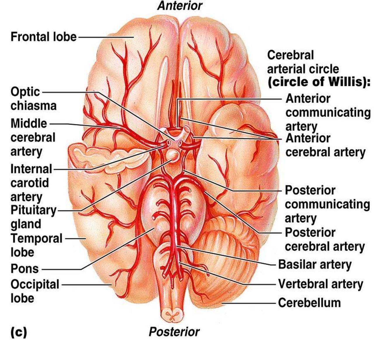
Pictures Of Cerebral Arteries from healthiack.com Explore the anatomy of the human cardiovascular system (also known as the circulatory system) with our detailed diagrams and information. The major deep veins of the arm are the radial and ulnar veins, which run along the length of their respective bones and merge at the elbow to form the. The common iliac arteries give off small branches to the psoas major, peritoneum, extraperitoneal. You can also use ohp permanent marker pens to label the structures after drawing them with thick i'm unsure if you're asking about general direction of flow or about memorizing specific names of major arteries and veins. Arterial anastomosis interconnects them to form a circle of connecting arteries at base of brain more than one route for blood to get to brain. Illustration depicting main leg arteries (anterior view). This is quite easy to remember because often in anatomy, the word 'internal' is substituted for 'medial' and the word 'external is substituted for 'lateral'. Anatomy of excitatory and conductive elements:
Major systemic arteries major systemic veins note:
Medial pectoral, lateral pectoral, intercostal, subcostal, phrenic, vagus, pelvic splanchnic. Roots, trunks, divisions, cords, branches. The major deep veins of the arm are the radial and ulnar veins, which run along the length of their respective bones and merge at the elbow to form the. So one would think there would be a pair of vertebral veins however, this is not the case in the brain. 15.1 abdominal aorta and major branches anterior view. Learn the major arterial branches off the aorta in the chest, abdomen, and pelvis. This artery stems from the external carotid artery, follows the inferior border of the mandible, and enters the face. This illustration was published in. Place the match each vein in column a with the vein it drains into from column b. Hansen, phd chapter:introduction to the human body page:14. Superficial vein collecting blood from the inner leg and thigh and receiving blood from certain veins of the foot; Place the letter of your choice in the space provided. Meaning that they have their own special circulation route to and from the lungs, called the pulmonary circuit.
Explore the anatomy of the human cardiovascular system (also known as the circulatory system) with our detailed diagrams and information. It is related to the femoral vein in the upper part of its course. Last updated on tue, 15 dec 2020 | human anatomy. Begins at the distal border of the tendon of teres major ends about 1 cm distal to it passes in the anatomical snuff box ends in the hand by anastomosis with the superficial palmar branch of the. Describe the waveforms and pressures that are seen in each anatomical location during insertion of a pulmonary artery catheter.
heart: Heart Veins And Arteries Labeled from image.slidesharecdn.com Major systemic arteries major systemic veins note: It is the longest vein in the body. You can also use ohp permanent marker pens to label the structures after drawing them with thick i'm unsure if you're asking about general direction of flow or about memorizing specific names of major arteries and veins. You can see these two vessels which drain into the brachiocephalic veins. The major vein collectors are integrated into the dura to form venous sinuses — not to be confused with. Hansen, phd chapter:introduction to the human body page:14. It is related to the femoral vein in the upper part of its course. Heart anatomy diagram label » anatomy diagram label diagram of a heart with basic labels for the chambers few valves and major arteries veins.
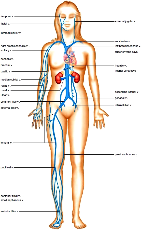
Arteries and veins of the human body.
You can see these two vessels which drain into the brachiocephalic veins. The femoral vein intervenes between the artery and the adductor longus. Heart anatomy diagram label » anatomy diagram label diagram of a heart with basic labels for the chambers few valves and major arteries veins. Roots, trunks, divisions, cords, branches. Learn anatomy faster and remember everything you learn. Related posts of anatomy of major veins and arteries. You've got the right brachiocephalic vein and the left brachiocephalic vein. Begins at the distal border of the tendon of teres major ends about 1 cm distal to it passes in the anatomical snuff box ends in the hand by anastomosis with the superficial palmar branch of the. The common iliac arteries give off small branches to the psoas major, peritoneum, extraperitoneal. There are three major types of blood vessels: Superior vena cava, azygos, hemiazygos, iliac veins, inferior vena cava nerves: This artery stems from the external carotid artery, follows the inferior border of the mandible, and enters the face. Superficial vein collecting blood from the inner leg and thigh and receiving blood from certain veins of the foot;
This illustration was published in. Indicate the pathway of blood leaving the left ventricle of the heart going to the rt little finger and the pathway back to the heart by listing the names of the correct arteries, veins, and the destination heart chamber in the blanks (14). Pulmonary arteries and veins function differently. Usually arteries and veins run together as they supply and drain specific areas of the body. The femoral vein intervenes between the artery and the adductor longus.
Circulatory Routes. Major Systemic Arteries. Major ... from encyclopedia.lubopitko-bg.com See the back for a diagram showing the two circulation routes. Roots, trunks, divisions, cords, branches. Place the match each vein in column a with the vein it drains into from column b. Superficial vein collecting blood from the inner leg and thigh and receiving blood from certain veins of the foot; Major systemic arteries major systemic veins note: Last updated on tue, 15 dec 2020 | human anatomy. Arteries and veins of the human body. Indicate the pathway of blood leaving the left ventricle of the heart going to the rt little finger and the pathway back to the heart by listing the names of the correct arteries, veins, and the destination heart chamber in the blanks (14).
Neither the pulmonary artery or vein are listed because they are not systemic;
Superficial vein collecting blood from the inner leg and thigh and receiving blood from certain veins of the foot; This artery stems from the external carotid artery, follows the inferior border of the mandible, and enters the face. Usually arteries and veins run together as they supply and drain specific areas of the body. Describe the waveforms and pressures that are seen in each anatomical location during insertion of a pulmonary artery catheter. Anatomy and physiology questions and answers. Learn anatomy faster and remember everything you learn. As they approach that organ they are usually accompanied by lymphatic vessels though not as a rule by arteries, and, sooner or later, they empty their blood into the deep veins. The common iliac arteries give off small branches to the psoas major, peritoneum, extraperitoneal. This illustration was published in. There are three major types of blood vessels: Learn the major arterial branches off the aorta in the chest, abdomen, and pelvis. Roots, trunks, divisions, cords, branches. It is related to the femoral vein in the upper part of its course.


0 Comments