Anatomy Of Chest Bones : Fractures of the bones in the chest, including ribs, sternum, clavicle, scapula and vertebrae.. When a patient flexes the neck forward, the prominent process is usually that of the 7th cervical. The scapula, or shoulder blade, is an approximately triangular shaped bone. Have you ever seen fossil remains of dinosaur and ancient human bones in textbooks, television, or in person at a museum? This anatomical midline can be useful in assessing for symmetry in breast augmentation or in performing a median sternotomy. Long bones are mostly located in the appendicular skeleton and include bones in the lower limbs (the tibia, fibula, femur, metatarsals, and phalanges) and bones in the upper limbs (the humerus, radius, ulna, metacarpals.
Bone of chest and their parts. Chest bone, ribs, lung, heart, xiphoid process. They are always longer than they are wide the vertebrae are irregular bones. It can help you understand our world more detailed and specific. Bone structure on plain studio background.human anatomy chest from low angle.
Bones of the Arm and Hand | Interactive Anatomy Guide from www.innerbody.com Identify the following structures on the lateral chest radiograph: Have you ever seen fossil remains of dinosaur and ancient human bones in textbooks, television, or in person at a museum? Compare the nuclear medicine scans to anatomical diagrams. They are always longer than they are wide the vertebrae are irregular bones. Right upper anatomy is to physiology as geography is to history: The largest bone in the human body is the thighbone or femur, and the smallest is the stapes in the middle ear, which are just 3 millimeters (mm) long. Long bones are categorised by their tubular shaft (diaphysis) with a rounded end (epiphysis) on each end. What can you label/identify on the nmt exam.
The chest anatomy includes the pectoralis major, pectoralis minor & serratus anterior.
Long bones are mostly located in the appendicular skeleton and include bones in the lower limbs (the tibia, fibula, femur, metatarsals, and phalanges) and bones in the upper limbs (the humerus, radius, ulna, metacarpals. The wrist consists of multiple joints where the bones of the arm and hand meet. Anatomy is the amazing science. Learn about each muscle, their locations & functional anatomy. Bone structure on plain studio background.human anatomy chest from low angle. Bones are mostly made of the protein collagen , which forms a soft framework. The mineral calcium phosphate hardens this framework, giving it. Right upper anatomy is to physiology as geography is to history: It, essentially, floats off of the back of the chest, as it is connected to the body primarily by muscle. Fractures of the bones in the chest, including ribs, sternum, clavicle, scapula and vertebrae. The largest bone in the human body is the thighbone or femur, and the smallest is the stapes in the middle ear, which are just 3 millimeters (mm) long. It originates at your clavicle, ribs, and sternum, and inserts into the upper portion of your humerus (upper arm bone from elbow to shoulder.) Computerized tomography 4 anatomy of lung segmental anatomy of lung lateral view on a normal lateral view the contours of the heart are visible and the ivc is seen entering the right atrium.
Learn about chest anatomy with free interactive flashcards. The ribs meet at an acute angle at the sternum, the costal cartilages thicken like beads at points of their transition to bones (rachitic beads). These bones form by the thickening of a. What can you label/identify on the nmt exam. Atlas of anatomy of the human body:
Rib Cage Stock Illustrations and Cartoons | Getty Images from media.gettyimages.com Long bones function to support the weight of the body and facilitate movement. Bone of chest and their parts. Ground substance and collagen fibers create a matrix that contains. An overview of the anatomy of the hand, including the bones of the hand, muscles, blood supply and nerve supply. Bones are mostly made of the protein collagen , which forms a soft framework. The manubrium, sternal body, and xiphoid process. Atlas of wrist mri anatomy. Computerized tomography 4 anatomy of lung segmental anatomy of lung lateral view on a normal lateral view the contours of the heart are visible and the ivc is seen entering the right atrium.
This is an important landmark, as the second costal cartilage is attached to it laterally, and.
Bone structure on plain studio background.human anatomy chest from low angle. Your rib cage, for example, acts like a shield around your chest to protect important organs inside such as your lungs and heart. Spot views were taken of the chest, spine, hand, and foot. This framework consists of many individual bones and cartilages. The largest bone in the human body is the thighbone or femur, and the smallest is the stapes in the middle ear, which are just 3 millimeters (mm) long. Atlas of wrist mri anatomy. These joints fuse together in adulthood, thus permitting brain growth during adolescence. The two bones are joined at a slight angle that protrudes anteriorly (sternal angle, angle of louis). Top suggestions for anatomy of chest bones. What can you label/identify on the nmt exam. Language and terminology for the study of the anatomy of the thorax. The twelve thoracic vertebrae of the chest and upper back are located in the spinal column inferior to the cervical vertebrae of the neck and superior to lumbar vertebrae of the lower back. Right upper anatomy is to physiology as geography is to history:
Spot views were taken of the chest, spine, hand, and foot. Anatomy is the amazing science. This anatomical midline can be useful in assessing for symmetry in breast augmentation or in performing a median sternotomy. It is comprised of many bones, formed by intramembranous ossification, which are joined together by sutures (fibrous joints). An overview of the anatomy of the hand, including the bones of the hand, muscles, blood supply and nerve supply.
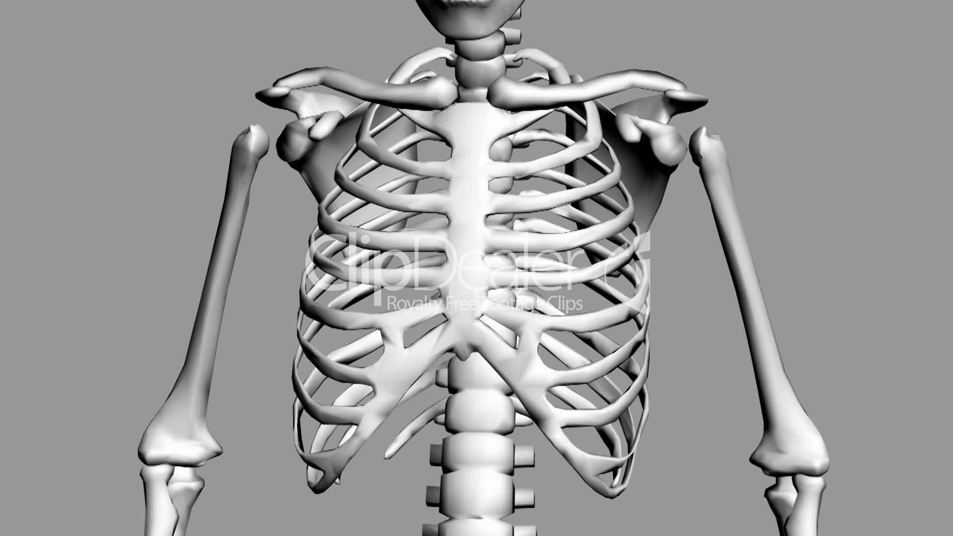
Rotation of 3D skeleton.ribs,chest,anatomy,human,medical ... from buidln.clipdealer.com Language and terminology for the study of the anatomy of the thorax. Your rib cage, for example, acts like a shield around your chest to protect important organs inside such as your lungs and heart. What can you label/identify on the nmt exam. Bones of the chest and upper back (posterior view). This webpage presents the anatomical structures found on wrist mri. We hope you will use this picture in the study and helping chest and abdominal cavities with some organs removed. This framework consists of many individual bones and cartilages. Human chest bone structure parts of the chest bones.
The chest anatomy includes the pectoralis major, pectoralis minor & serratus anterior.
When a patient flexes the neck forward, the prominent process is usually that of the 7th cervical. Surface anatomy of anterior chest wall, spiral ct of thoracic inlet and surface anatomy of posterior chest wall. Your rib cage, for example, acts like a shield around your chest to protect important organs inside such as your lungs and heart. Bones are mostly made of the protein collagen , which forms a soft framework. The ribs and sternum make up what is called the ribcage the ribcage protects the lungs blood vessels and heart along with parts of the sple. The ribs meet at an acute angle at the sternum, the costal cartilages thicken like beads at points of their transition to bones (rachitic beads). Bone basics and bone anatomy. Learn about chest anatomy with free interactive flashcards. It describes the theatre of events. Bones have many shapes and sizes and are important to add structure to the body and protection to the vital structures. There also are bands of fibrous connective tissue—the ligaments and the tendons—in intimate relationship with the parts of the skeleton. Spot views were taken of the chest, spine, hand, and foot. What can you label/identify on the nmt exam.
Sesamoid bones are generally small, flat and have an apex at one end anatomy of chest. Long bones function to support the weight of the body and facilitate movement.


0 Comments