Diagram Of Bones In Neck And Shoulder - Sensors Free Full Text A Two Joint Neck Model To Identify Malposition Of The Head Relative To The Thorax Html - In the front of the neck, the platysma muscle extends up from the chest, goes over the.
Diagram Of Bones In Neck And Shoulder - Sensors Free Full Text A Two Joint Neck Model To Identify Malposition Of The Head Relative To The Thorax Html - In the front of the neck, the platysma muscle extends up from the chest, goes over the.. It consists of two major parts: Contains glands ( thyroid, parathyroid, and thymus ), the larynx, pharynx and trachea. Shoulder girdle an overview sciencedirect topics. These bones have some interesting landmarks, including various bumps and projections. The neck and shoulders are complex and interconnected areas, and medical problems that affect one often affect the other, as well.
The upper arm bone, called the humerus, is connected to the body via the shoulder blade, which possesses the latin name scapula. Contains cervical vertebrae and postural muscles. There are seven of them. Where the rounded top of the arm bone (humerus) contacts the shoulder blade is. The neurocranium is the part enveloping the brain and is formed out of two parts;
Upper Cervical Spine Disorders Anatomy Of The Head And Upper Neck from www.spineuniverse.com 2.1 bones of the shoulder girdle 2.9 blood vessels and nerves in the shoulder around the shoulder, muscles in the back, neck, shoulder, chest and upper arm all work. However, the muscles of the neck can also be easily strained or injured. The content of the neck is grouped into 4 neck spaces, called the compartments. The neck and shoulders are complex and interconnected areas, and medical problems that affect one often affect the other, as well. Shoulder joint of human body anatomy infographic diagram with all parts including bones ligaments muscles bursa cavity capsule cartilage membrane for medical science. The human head and neck bones are crucial for structure and support. With one on each side of the neck, these help flex the. Degenerative arthritis of the spine in the neck (cervical spine) can pinch nerves that can cause both neck pain and shoulder pain.
Cervical spine anatomy is quite complex.
The skull is a strong, bony capsule that rests on the neck and encloses the brain. Diagram of bones in neck and shoulder. The muscles of the neck provide the mechanism for swallowing, yawning, speaking, and moving the head. The ligaments connect the three bones — the humerus, or upper arm bone; However, the muscles of the neck can also be easily strained or injured. The upper arm bone, called the humerus, is connected to the body via the shoulder blade, which possesses the latin name scapula. Contains cervical vertebrae and postural muscles. This may cause pain that radiates into the shoulder, as well as numbness that travels down the arm and into the hand. The neck and shoulders are complex and interconnected areas, and medical problems that affect one often affect the other, as well. The neck bones are called the cervical vertebrae. Innerbody research is the largest home health and wellness guide online, helping over one million visitors each month learn about health products and services. Axial skeleton — bones of the skull, vertebral column, thoracic cage. The scapula, or shoulder blade;
The skull is a strong, bony capsule that rests on the neck and encloses the brain. In adults the long bones of the legs and arms are filled with yellow marrow. Degenerative arthritis of the spine in the neck (cervical spine) can pinch nerves that can cause both neck pain and shoulder pain. To ensure proper range of motion, the shoulder joint is supported by the shoulder ligaments, shoulder tendons and shoulder muscles. It also provides sensation to parts of the upper arm.
Labeled Anatomy Chart Of Triceps Muscles Isolated In Skeleton On White Background Stock Photo Download Image Now Istock from media.istockphoto.com Cervical radiculopathy, commonly called a pinched nerve occurs when a nerve in the neck is compressed or irritated where it branches away from the spinal cord. In this video part, you will also find out the anatomy of the neck and shoulders. Shoulder skeleton diagram with head and deltoid tubercle of humerus, scapula skeletal structure anatomy of neck and shoulder stock illustrations The shoulder tendons attach to the bones at one. Innerbody research is the largest home health and wellness guide online, helping over one million visitors each month learn about health products and services. Where the rounded top of the arm bone (humerus) contacts the shoulder blade is. The three bones of the shoulder are the: The bones of the head and neck play the vital role of supporting the brain, sensory organs, nerves, and blood vessels of the head and protecting these structures from mechanical damage.
This is called the glenoid.
Diagram of bones in neck and shoulder. Cervical osteoarthritis (neck arthritis) osteoarthritis occurs when the protective cartilage in a joint begins to break down and no longer facilitates smooth movement between bones, which can eventually result in the joint becoming swollen and painful. The cervical spine, your neck, is a complex structure making up the first region of the spinal column starting immediately below the skull and ending at the first thoracic vertebra. Contains cervical vertebrae and postural muscles. However, the muscles of the neck can also be easily strained or injured. The bones of the head and neck play the vital role of supporting the brain, sensory organs, nerves, and blood vessels of the head and protecting these structures from mechanical damage. Pain and dysfunction from injuries or conditions that impact the joints, muscles, and other structures can easily spread from the neck to the shoulder(s) and from the shoulder(s) to the neck. Degenerative arthritis of the spine in the neck (cervical spine) can pinch nerves that can cause both neck pain and shoulder pain. The skull base that supports the brain and the calvaria (skullcap) that sits on top of the base, covering the brain. The muscles of the neck provide the mechanism for swallowing, yawning, speaking, and moving the head. It consists of two major parts: This may cause pain that radiates into the shoulder, as well as numbness that travels down the arm and into the hand. Contain the common carotid artery, internal.
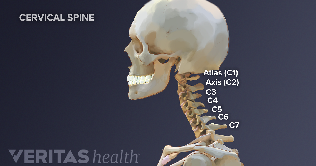
Although anchored in the neck, their primary functions are to move the shoulder blades and support the arms. Bone diagram forehead (frontal bone) nose bones (nasals) cheek bone (zygoma) upper jaw (maxilla) lower jaw (mandible) breast bone (sternum) upper arm bone (humerus) lower arm bone (ulna) thigh bone (femur) collar bone (clavicle) toe bones (phalanges) ankle bones (tarsals) kneecap (patella) shin bone (tibia) calf bone (fibula) foot bones Cervical spine anatomy is quite complex. C6 is the nerve root that exits the spinal cord above the sixth vertebra in the neck. 2.1 bones of the shoulder girdle 2.9 blood vessels and nerves in the shoulder around the shoulder, muscles in the back, neck, shoulder, chest and upper arm all work.
Cervical Spine Anatomy from embed.widencdn.net Bone diagram forehead (frontal bone) nose bones (nasals) cheek bone (zygoma) upper jaw (maxilla) lower jaw (mandible) breast bone (sternum) upper arm bone (humerus) lower arm bone (ulna) thigh bone (femur) collar bone (clavicle) toe bones (phalanges) ankle bones (tarsals) kneecap (patella) shin bone (tibia) calf bone (fibula) foot bones The neurocranium is the part enveloping the brain and is formed out of two parts; With the weight that must be supported, these muscles are strong. There are seven cervical vertebrae that allow for a great amount of motion in the neck. Boneka stitch jumbo warna biru. There are seven of them. The shoulder tendons attach to the bones at one. The ligaments connect the three bones — the humerus, or upper arm bone;
Contains glands ( thyroid, parathyroid, and thymus ), the larynx, pharynx and trachea.
This may cause pain that radiates into the shoulder, as well as numbness that travels down the arm and into the hand. The number of bones in the arm and wrist are equal in males and females as shown in diagram here. Although anchored in the neck, their primary functions are to move the shoulder blades and support the arms. It consists of two major parts: In adults the long bones of the legs and arms are filled with yellow marrow. Axial skeleton — bones of the skull, vertebral column, thoracic cage. A second joint in the shoulder is the junction of the collar bone with the shoulder blade, called. Bone diagram forehead (frontal bone) nose bones (nasals) cheek bone (zygoma) upper jaw (maxilla) lower jaw (mandible) breast bone (sternum) upper arm bone (humerus) lower arm bone (ulna) thigh bone (femur) collar bone (clavicle) toe bones (phalanges) ankle bones (tarsals) kneecap (patella) shin bone (tibia) calf bone (fibula) foot bones The first one that holds the skull is called the atlas. The neurocranium is the part enveloping the brain and is formed out of two parts; Bones of the head and neck. The bones of the head and neck play the vital role of supporting the brain, sensory organs, nerves, and blood vessels of the head and protecting these structures from mechanical damage. Each arm is attached to a shoulder blade.


0 Comments